
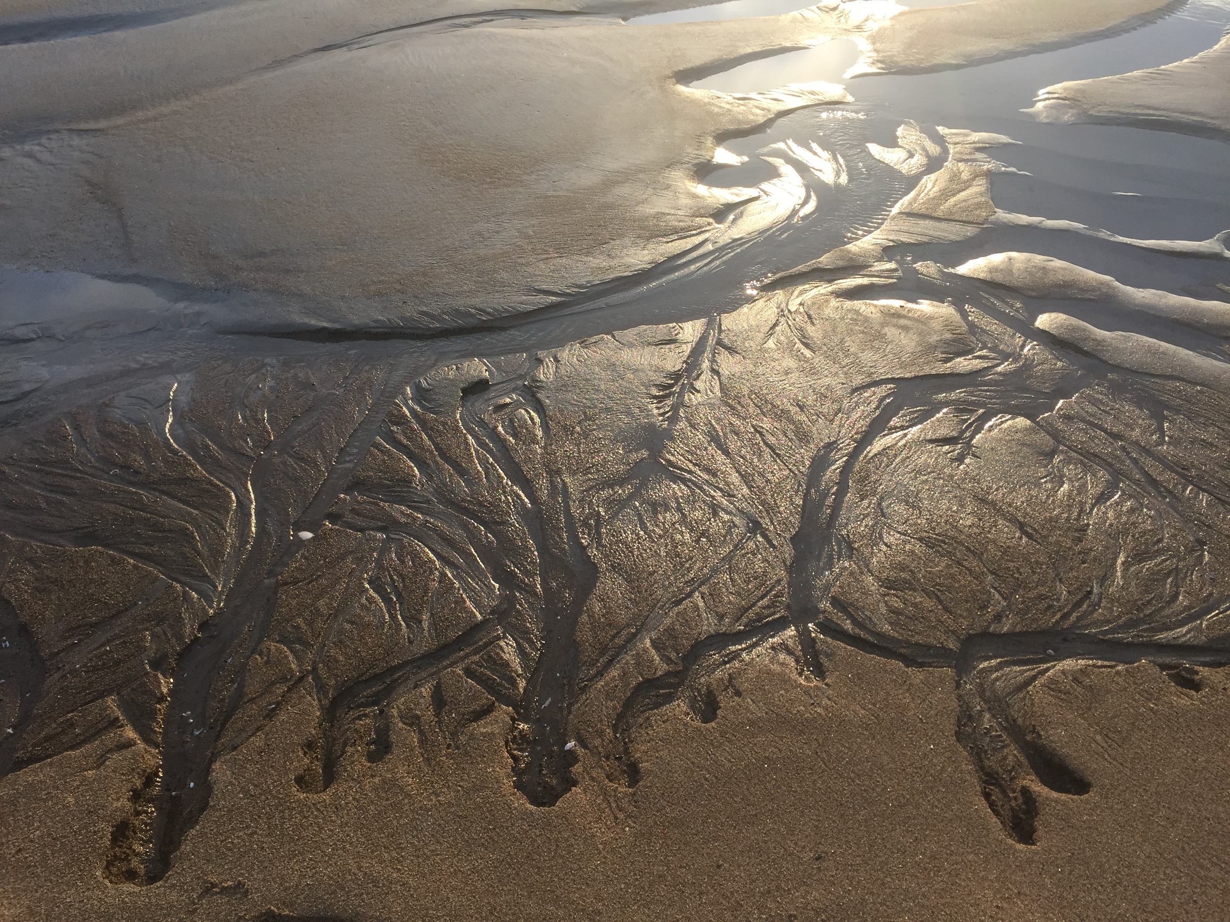


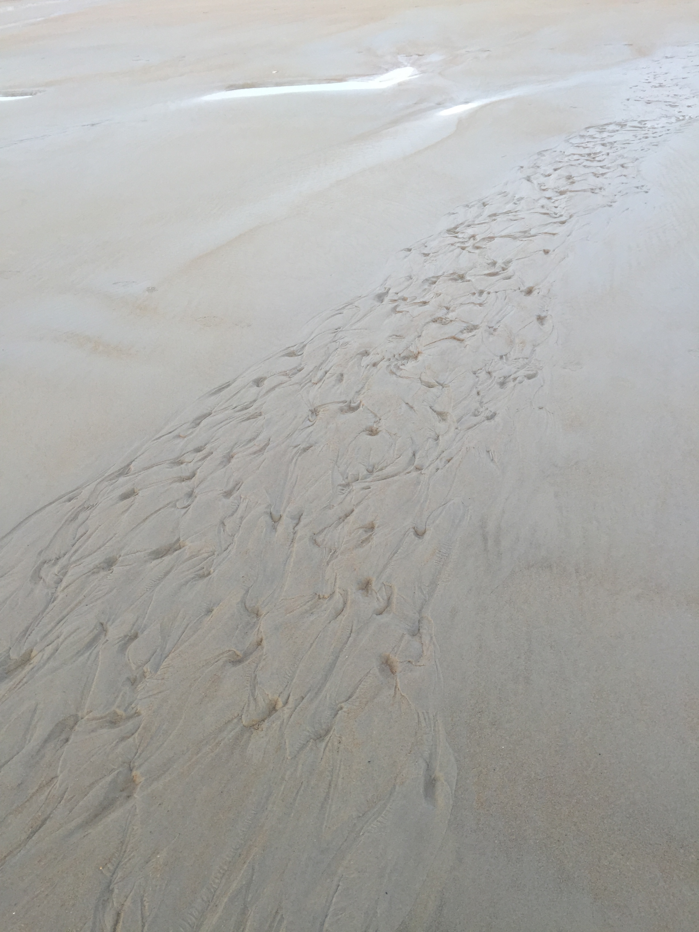
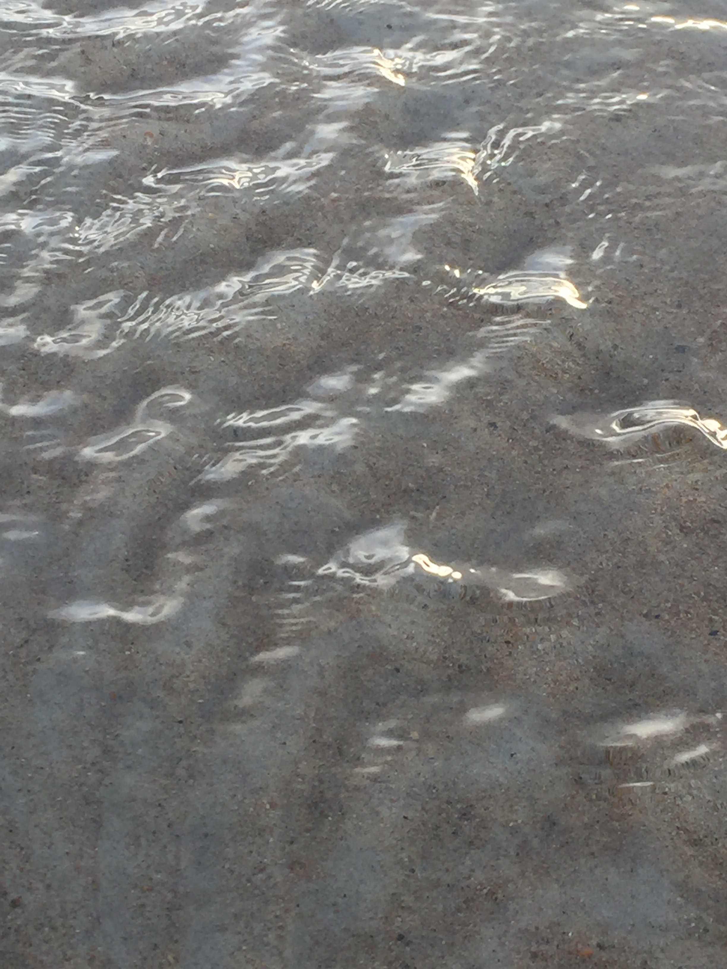
“Notice all around you - flow - circulation - transformation”


Homework and Preparation Fluids 2016
Prepare your Anatomy:
Review and color the sections covering these fluids in the Anatomy Coloring Book- pages CSF 141-143, circulation 81, 82, Blood 100-118 , especially 100-102,106-108 and 111-113, 115,118, Lymph 120,125( if you prefer to make models or free hand draw all this instead of coloring - please do!)
For Synovial see synovial handout, for 3D Spiral Blood flow here, lymphatic system here, Synovium is a connective tissue membrane that lines the cavity between joints and secretes a lubricating fluid known as the synovial fluid., Arteries and Veins slide show, lub-dub of heart, Interstitial or Extracellular fluid here
Watch these youtube videos: Heart Flow here, or slower here, the helical heart here, Urinary System here, more info on Urinary here, and here, Cerebrospinal fluid here, then add this info, and here, , more on Meninges here, and Cerebral veins here, and here, wanna get real detailed and draw? ( not necessary , but may explain some unclear transitions)
Create a fluid Model: demonstrating one of the Fluid Systems of the body: Circulation, Lymph, Cellular, Cranial Sacral, Interstitial, Synovial or make a fluid or gel game toy video, audio podcast, a thatre peice a treasure hunt a closed eye experience a tasting club .......
Explore your fluids - either in dance or in a regular practice you do: dish washing, yoga, washing a baby, hiking, biking, etc.
Stay connected with your folks!
through all this - follow your curiosity, make your own way, add in things that interest you........
Links to emails (on google docs)
email #2 pathway of the fluids - vasculature
email #3 deeper into fluids
email #4 fluids and muscles
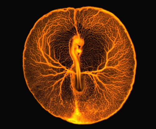
What does this image show?
The image shows the central chicken embryo surrounded by the vitelline vascular system, the veins and arteries supporting the flow of blood between the embryo and the yolk sac. The head of the embryo, including the embryonic eye and brain, can be seen on the upper part of the embryo, just above the embryonic heart. The long lower part of the embryo is the future body of the chicken, from which legs and wings will develop.
How was this image acquired?
The image was obtained by opening the egg's shell and injecting a fluorescent dye into the vascular system (the lower part). Because of the action of the pumping heart, the dye spread to the entire vascular system (vitelline veins and arteries). Images were then recorded using a fluorescence microscope equipped with a camera. The image shows the whole embryo as a result of monitoring total reflected fluorescence. http://www.wellcomeimageawards.org/2012/chicken-embryo-vascular-system
Oh WAIT - this is also crazy amazing - really - sorry I'm so over the top, but: check it out! Hint: - Its 3D
Brooke Thomas' Liberated Body podcast