Images
Excerpt from a workshop description by Nicole Binder, BMC practitioner:
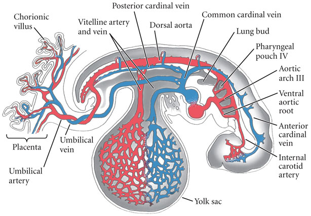
......The arterial blood swirls out toward the distal parts of the body and has a repetitive, pulsing rhythm. The venous blood whorls back toward the heart and has a wavelike rhythm. The place where the arterial and venous capillaries meet is called the isoring, a resting place between coming and going. Through balancing this inward and outward flow and finding the meeting place of stillness, we can move with more facility between solo dancing, meeting others, and sharing weight.
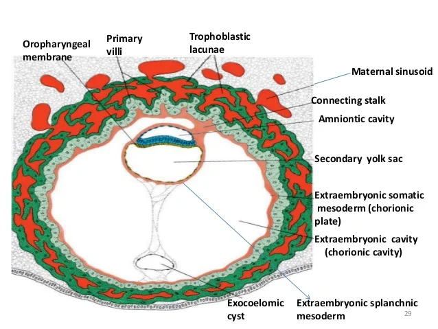
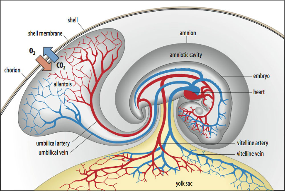
This workshop will also explore polarities as they manifest in the blood and cerebrospinal fluid, which are the heaviest and lightest fluids in the body. In utero the blood forms in the yolk sac in the front, and the cerebrospinal fluid arises from the amniotic fluid in the back. Within these two fluids we can find opposition and balance between front / back, inner / outer, gravity / space, folding / extending, rolling / sloughing, and underdancer / overdancer. The transitions between these poles are nonlinear and take us into spiral pathways......
Videos
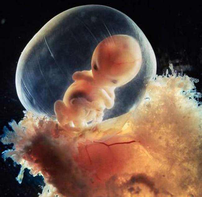
1st video - watch without sound and notice how the outer tissues of the embryo relate to the uterine wall
2nd video - at 3:40 you can see how fast the new heart is pumping!
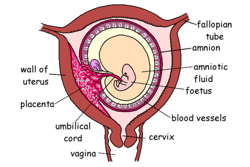
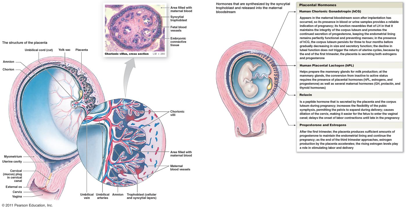
Placenta video - midwife shows how the amniotic sac connects to the placenta - she is also just so relaxed!!
4th video might inspire you to draw!!
8 Medical Embryology - go to 13:25- 15:10 it shows the blood islands in the arch that becomes that heart that have not fused yet! then 17:40 on it shows the heart tube shifting to bend and wrap around itself. Don't worry about all the other details!!
EMBRYO ONLINE Resources
WELCOME to the Online Embryo Course.
You are ALWAYS welcome to share a video, drawing , writing, feelings, either directly to Christine - embodyourlife@gmail.com or to the facebook group or the email list. facebook page here
Online Embryology Sources: Free Online Embryology- simple, Free Online very detailed embryology, Blechschmidt models
Audio and Text
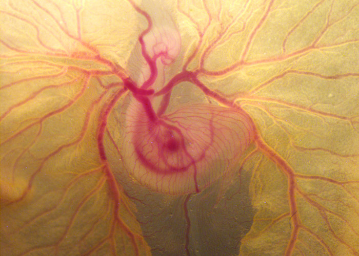
Watch out for how the embryo's system creates blood vessels, then connects these into veins and arteries to harvest nutrients from the tissues close to the mother's capillaries.
Then notice that it establishes blood vessels to feed on its own yolk sac, transforming this nourishment into cells, organs, tissues of its new body.
Click on the LEFT TOP BARS on the video screen to see the complete PLAYLIST

